Microscopes are designed to image flat surfaces, unlike the curved structures typical for biology. Here, I would like to mention a piece of work where we demonstrate a method based on K-mirrors to overcome this lack of flexibility for two-photon resonant scanning microscopes.
In my work, I regularly use two-photon microscopy to image the hippocampal layer of pyramidal cells in mouse CA1. For years, I noticed again and again that the cell layer I wanted to image was sometimes captured perfectly (as shown in typical published figures), but more often cut through by the imaging plane at an arbitrary angle, as shown in the two examples below, leaving a diagonal band of cells (imaged by Shinjini Ghosh).
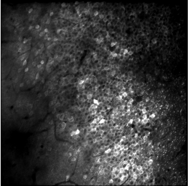
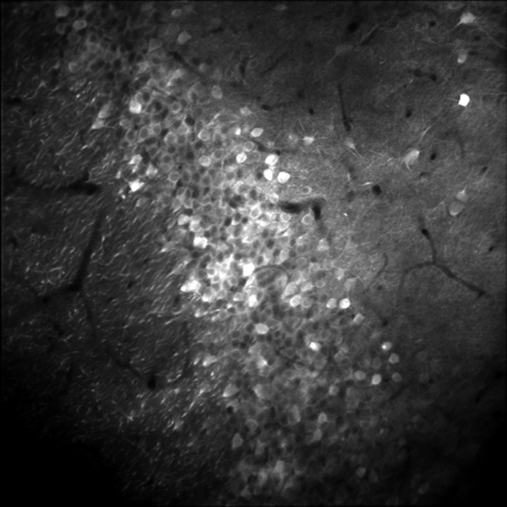
This is an issue that most neuroscientists using a two-photon microscope may have encountered. It is quite prominent for mouse CA1, but it is even more striking for mouse CA2 or CA3. And I was struggling with similar issues even when I worked on the small and curved brain of the adult or larval zebrafish during my PhD.
In principle, there is a simple solution to this problem: you can simply curve and tilt the imaging plane, using a fast z-scanning device such as a tunable lens or a remote mirror (which I implemented 10 years ago). However, these “fast” z-scanning devices are still slow in the range of several milliseconds settling time. This makes it possible to change the z-focus for the slow y-axis (typically 30 ms per scan) but not for the fast x-axis (65 μs per scan). Therefore, you cannot curve and tilt the image plane along the direction that would be ideal.
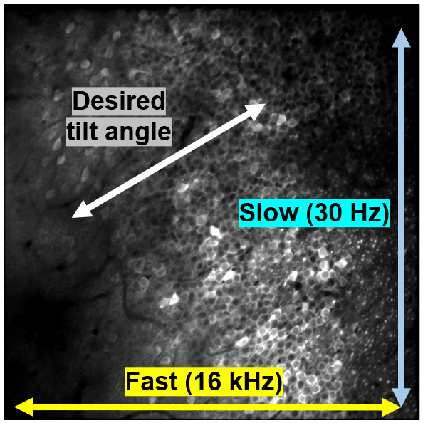
In 2024, the solution to this problem hit me in the face: a very simple but effective solution. I read this tweet by microscopy expert Reto Fiolka, in which they used galvo scan mirrors to build a fast SIM microscope. It took me a bit of time to understand what they were doing here, but then I realized that the principle they were describing – the use of K-mirrors to rotate a FOV – was not only the solution to my problem, but also an established method in astronomy for perhaps 70-80 years!
With my fearless PhD student, Shinjini Ghosh, we designed a simple add-on to resonant scanning two-photon microscopes that performs this FOV rotation and thus can optimize for example the imaging of pyramidal cells in hippocampal CA1.
Check out our preprint, where we demonstrate therange of applications that benefit from this small modification, ranging from CA1 pyramidal cells, hippocampal dendrites, cortical layers, prism-based dendritic imaging, cerebellar Purkinje cell imaging, up to optimized glutamate imaging. Here’s an example, where we have a tilted FOV in hippocampal CA2/3, where we demonstrate that FOV rotation with the K-mirror, combined with axial scanning can be used to bring the hippocampal dendrites beautifully into the FOV:
For many more applications, and a thorough documentation of the optical design, check out our preprint (https://doi.org/10.64898/2026.10.09.757907) and our GitHub repository!


Greetings, Peter. In regard to rotating the field from a very different angle\ pun intended, I am involved in capturing a 3D Modeling bones in a cave without getting close.\not Touching Anything in the proximity… We are using lidar, yet. Wanted to get more detail. Briefly reading it not totally understanding it did remind me of our situation and\n I wondered if you might capture the whole in 3D using new methods.https://share.google/g01srFnDGNezhBE52 Came across this material and thought it might give you some other ideas to add to your own…. Hey, you never know about these things.All the best, jim Leishman,that sculptor guy back in Canada