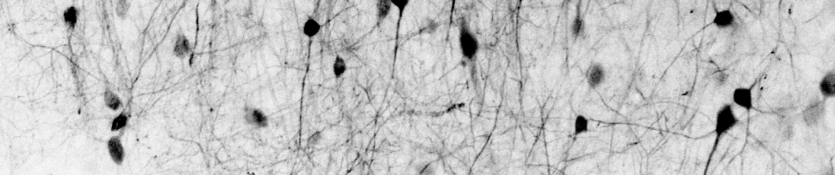
Few things in neuroscience are more beautiful than the ultrastructure of individual neurons. Zhihao Zheng and his colleagues from the Tank/Seung labs have now published a 3D electron microscopy dataset from hippocampal CA3.
All neurons in this volume were automatically segmented and proofread. But what about glia? The paper only briefly mentions “non-neuronal” cells and does not consider them in the analysis. However, the automated segmentation typically also produces – as byproducts – segmented astrocytes or other glia cells (although probably not proofread and therefore less trustworthy). I looked into the publicly accessible dataset and soon found some putative glial cells.
Here’s an interesting pair of cells: a pyramidal neuron with its large cell body sitting on top of what may be a microglia cell (alternatively, but less likely, an interneuron). I have no idea why these two cell bodies are so close to each other. If you have an idea, let me know! (Here’s the link to look at this cell pair yourself.)
Next, I specifically looked out for astrocytes, which are currently my favorite brain cell type. Since these cells are most likely not carefully proofread, the reconstructions need to be taken with a grain of salt. Nevertheless, the difference compared to morphologies of pyramidal neurons is really striking: astrocytes form much denser and local branches. They appear like sponges rather than stars, at least fromthis ultrastructural perspective. The video is slow and not smooth because the many astrocytic branches caused lag during my screen recording. Here’s the link to look at these astrocytes yourself.
For the blue-colored astrocyte, you can even see a putative endfoot that wraps around a blood vessel (although this would need to be proofread to be sure). In addition, it is possible to directly see the dense coverage of space by astrocytic processes – again with the caveat that these astrocytic segmentations probably have not be proofread.
It’s always interesting to explore 3D electron microscopy datasets. This one is not of the highest resolution (45 nm in the z-axis), but it is definitely fascinating to explore if you are interested in hippocampal circuits, be it from the perspective of neurons or astrocytes!


