Publications

Check out my Google Scholar profile for a more complete publication list. My PhD thesis can be found online as a pdf.
miniML: A deep learning framework for synaptic event detectionRupprecht P, Rozsa M, Fang X, Svoboda K, Helmchen F (2025). Spike inference from calcium imaging data acquired with GCaMP8 indicators. bioRxiv (2025) Preprint!
This work is a comprehensive analysis of spike inference with the new calcium indicator variants GCaMP8. The study addresses the following questions: How does GCaMP8 differ from previous generations like GCaMP6? Can it detect single spikes better or not? Can models for spike inference trained on GCaMP6 be directly applied to GCaMP8? Which parameters should I choose to optimize spike inference with GCaMP8 data with MLSpike or Suite2p (OASIS)? The most interesting section, in my opinion, is however Figure 3 on the effect of the linearity of a calcium indicator on how complex spike patterns are seen with calcium imaging, showing systematic changes between GCaMP6 (more non-linear) and GCaMP8 (more linear) variants (illustrated to the left). I believe that anybody interested in calcium imaging can find something interesting or even many interesting aspects in this manuscript. I’m also proud to say that the paper includes an analysis that is based on the thesis of the first Master’s student of my independent research group, Xusheng Fang – I’m very happy about his contribution!
Centripetal propagation of calcium signals from distal to somatic compartments in astrocytes, triggered by optogenetic stimulation of the locus coeruleusRupprecht P, Wei F, Sullivan SJ, Helmchen F, Sdrulla AD. Spike rate inference from mouse spinal cord calcium imaging data. Journal of Neuroscience (2025).
CASCADE is a toolbox for spike inference that I maintain and improve continuously. In Spring 2023, I was approached by Andrei Sdrulla from OHSU in Portland. They wanted to test CASCADE for their calcium imaging data from spinal cord. In addition, they had ground truth recordings of simultaneously recorded calcium and electrophysiological data from the same neurons in spinal cord. I realized that this dataset was useful and interesting, not only for this specific project but also for anybody doing calcium imaging in spinal cord. Therefore, I performed systematic and detailed analyses that show how spike inference generalizes from cortex data to spinal cord. These analyses are not only interesting for spinal cord experts but also for those who wonder how calcium indicators and neurons behave similarly or differently across regions of the CNS. For example, Figure 2 nicely contrasts the different bursting behaviors of spinal cord vs. cortical neurons. And Figure 3 is, in my opinion, a very intuitive illustration of calcium transients evoked by single spikes in spinal cord vs. cortical neurons.
miniML: A deep learning framework for synaptic event detectionO’Neill PS, Baccino-Calace M, Rupprecht P, Lee S, Hao YA, Lin MZ; Friedrich RW, Mueller M, Delvendahl I. A deep learning framework for automated and generalized synaptic event analysis. eLife 13 (2025).
This is work mostly done by Philipp O’Neill and Igor Delvendahl. They developed miniML, an algorithm to detect single synaptic events from whole-cell patch-clamp data. Working in the same building, we talked about the algorithm, and I was soon convinced by the consistent quality of event detection and the clarity of their analyses. During my PhD, I had myself recorded whole-cell recordings in an explant zebrafish brain (published here). These data presented themselves as an interesting challenge to miniML because of the high frequency of synaptic events and the large diversity of event shapes. My contribution to this study was to analyze the events detected by miniML for this dataset and to describe what can be learnt from the detected events (Fig. 6). If you are into whole-cell synaptic events, in particular the analysis of “minis”, check out this excellent method (also on Github)!
A distinct hypothalamus-habenula circuit governs risk preference
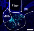
Groos D, Reuss AM*, Rupprecht P*, Stachniak T, Lewis C, Han S, Roggenbach A, Sturman O, Sych Y, Wieckhorst M, Bohacek J, Karayannis T, Aguzzi A, Helmchen F. A distinct hypothalamus-habenula circuit governs risk preference. Nature Neuroscience (2025)
In this tour-de-force PhD project, Dominik Groos investigated the role of the lateral habenula (LHb) during decision-making. The LHb is thought to be mostly involved in negative emotional states and depression, but Dominik found that neurons in the LHb also represent choice preference. He and his team used an array of methods, ranging from fiber photometry, 2P GRIN lens imaging, light-sheet imaging, optogenetics, retrograde tracing, slice patching etc. to dissect this circuit motif projecting from different hypothalamic nuclei to the LHb. My contribution to this huge project was the analysis of a part of the behavioral data, the construction of the two-photon microscope and the setting up of the two-photon analysis pipeline for GRIN lens imaging.
Centripetal propagation of calcium signals from distal to somatic compartments in astrocytes, triggered by optogenetic stimulation of the locus coeruleusRupprecht P, Duss SN, Becker D, Lewis CM, Bohacek J, Helmchen F. Centripetal integration of past events by hippocampal astrocytes regulated by the locus coeruleus. Nature Neuroscience (2024)
In this main project of my postdoc with Fritjof Helmchen, I found that astrocytic somata integrate calcium signals from distal processes on a slow timescale, but only if high arousal permits such a centripetal propagation of calcium signals. I believe that this might be a fundamental functional property of astrocytes in vivo. We found these specific spatio-temporal patterns during an exploratory analysis of the raw data based on some initial observations. Then, I used deep learning-based self-consistent denoising and applied correlation functions to each pixel’s time trace of the denoised movie. By doing so, I could compute the average delay of each pixel’s activity trace with respect to a reference signal (here: the global mean activity across the FOV), resulting in a delay map with short delays for processes and long delays for somata. For the revised version of the manuscript, together with the Bohacek Lab, we found that optogenetic stimulation of the locus coeruleus was sufficient to trigger this centripetal propagation (see the gif on the left side). Check out the paper or preprint for many more details and analyses!
Centripetal propagation of calcium signals from distal to somatic compartments in astrocytes, triggered by optogenetic stimulation of the locus coeruleusMasala N, Mittag M, Giovannetti EA, O’Neil DA, Distler F, Rupprecht P, Helmchen F, Yuste R, Fuhrmann M, Beck H, Wenzel M, Kelly T. Aberrant hippocampal Ca2+ micro-waves following synapsin-dependent adenoviral expression of Ca2+ indicators. eLife (2024)
The hippocampus is one of the most sensitive brain regions and is therefore often involved in pathologies such as epilepsy. Correspondingly, it seems relatively easy to perturb the natural state of hippocampus as a side-effect of experiments. Here, the team lead by Tony Kelly and Michael Wenzel describes pathological “micro-waves” that can be observed in the hippocampus but not cortex with calcium imaging and are most likely induced by high-titer and hSynapsin-dependent AAV expression of GECIs. My contribution to this study was to observe the same phenomenon also in a category of GECIs that is distinct from the jGCaMP family (2P imaging of CA1 with R-CaMP1.07, see gif on the left). An important study to keep in mind if you are doing calcium imaging in hippocampus.
Simultaneous calcium and spike recording in a zebrafish neuronRupprecht P, Carta S, Hoffmann A, Echizen M, Blot A, Kwan AC, Dan Y, Hofer SB, Kitamura K, Helmchen F*, and Friedrich RW*. A database and deep learning toolbox for noise-optimized, generalized spike inference from calcium imaging. Nature Neuroscience (2021)
In this large effort, we collected a database of almost 300 neurons with simultaneous calcium imaging and juxtacellular recordings of action potentials, across 8 calcium indicators and 9 brain regions in 2 species, with a total of almost 500,000 action potentials. I used this database to train a powerful algorithm based on deep networks to recover spike rates from calcium recordings, which generalized very well to unseen data. The algorithm uses its knowledge about the spike-calcium relationship and therefore is able to both recover spike rates at an improved temporal resolution and to suppress noise. Many more details and insights are in the paper, which could be a very interesting read to anybody interested in calcium imaging. Essential parts of this study are an easy-to-use Github repository that includes both code and the ground truth datasets, and a Colab Notebook that can be applied to your data, without installation or parameter tuning.
Four neurons in CA3 color-coded by their distinct activity patternsSchoenfeld G*, Carta S*, Rupprecht P, Ayaz A, Helmchen F. In vivo calcium imaging of CA3 pyramidal neuron populations in adult mouse hippocampus. eNeuro (2021)
This study was led by Gwendolin Schoenfeld, and I joined this project only at a later stage for data analysis, interpretation of results and writing. Two findings that I would like to highlight: First, despite reports that tuning of CA3 neurons is unstable across days, we found that patterns of co-activity remained stable across days; co-activity therefore might be conserved across days, while tuning is less so. Second, using simultaneous calcium imaging and juxtacellular recordings, we found a supra-linearity of calcium, hinting at the involvement of calcium spikes, a potential learning-related signal. We have performed these difficult juxtacellular experiments together with calcium imaging in anesthetized animals, but it would be really interesting to repeat this in awake animals.
Head-fixed adult zebrafishHuang KH, Rupprecht P, Frank T, Kawakami K, Bouwmeester T, Friedrich RW. A virtual reality system to analyze neural activity and behavior in adult zebrafish. Nature Methods (2020)
In this study, Kuo-Hua Huang developed a method to head-fixate adult zebrafish, made them interact with a virtual reality and imaged neuronal acitivty through the skull – all at the same time. I was mainly involved in the technical parts of the study and in the writing of the manuscript. Specifically, I helped to install a resonant scanning microscope system and to synchronize it via the control software (ScanimgeB) with the virtual reality. In addition, this was the first time when my and Stephan Gerhard’s algorithm for calcium signal deconvolution (Elephant) was really crucial, namely to reveal fast, swim-triggered dynamics of neuronal activity that are masked by the slow transients of calcium indicators. Overall, this is a really cool study and I’m proud to have contributed to this work. (Read-only PDF)
SpikefinderRupprecht P, Friedrich RW. Precise synaptic balance in the zebrafish homolog of olfactory cortex. Neuron (2018)
This is the main paper from my PhD thesis. Having developed methods to record and analyze calcium population data during the first part, I switched to whole-cell voltage clamp recordings in single neurons during the second part of my PhD. I was less interested in the coarse description of neuronal population activity, but rather in the mechanisms underlying the firing of single neurons. This work tries to understand how the biophysical properties of single neurons constrain or enable computational properties of the underlying circuit. Therefore, this experimental study directly connects to questions arising from theoretical neuroscience; in general, I think that intracellular electrophysiology in the intact brain  might be the best tool to test theoretical circuit models due to its high precision. The picture to the left shows a single neuron that is slowly filled with a dye by the micropipette after break-in. (PDF, SI)
SpikefinderBerens P, Freeman J, Deneux T, Chenkov N, McColgan T, Speiser A, Macke JH, Turaga S, Mineault P, Rupprecht P, Gerhard S, Friedrich RW, Friedrich J, Paninski L, Pachitariu M, Harris KD, Bolte B, Machado TA, Ringach D, Reimer J, Froudarakis E, Euler E, Roman-Roson M, Theis L, Tolias AS, Bethge M. Community-based benchmarking improves spike inference from two-photon calcium imaging data. PLoS Computational Biology (2018)
This paper is the result of the spikefinder competition, with the goal to solve the (inverse) problem of spike inference for calcium imaging data. I participated in the competition, together with Stephan Gerhard, using an algorithm based on 1D convolutional networks and embedding spaces, which got us a first prize. More details are on this blog (link 1, link 2, link 3), in the paper itself and on Github.
nice-ob-recording-6if-kept-stack-1Jacobson GJ, Rupprecht P, Friedrich RW. Experience-dependent plasticity of odor representations in the telencephalon of zebrafish. Current Biology (2017)
Most of the work and analysis in this paper was done by Gilad Jacobson. I joined the project when it came to recording the neuronal population activity of mitral cells in the olfactory bulb. Those cells are scattered in 3D, which makes it necessary to perform multi-plane calcium imaging to simultaneously record from a decent number of cells. I did these experiments, taking advantage of the voice coil-based remote z-scanning that I had developed before. Dynamics in the olfactory bulb are very rich and fascinating; mitral cells respond together with the large dendritic tuft (some 10 μms in diameter), which makes the visualization more fascinating than blinking somata alone (a small excerpt of a FOV is shown to the left).
Rupprecht P, Prendergast A, Wyart C, Friedrich RW. Remote z-scanning with a macroscopic voice coil motor for fast 3D multiphoton laser scanning microscopy. Biomedical Optics Express (2016)
This is one of the side-projects of my PhD. It lead to a method for fast 3D scanning for two-photon imaging which I have been using since routinely for multi-plane calcium imaging, replacing the more expensive and fragile standard technique (piezo-attached objectives). The design is described elsewhere on this website (link). For me, who was rather new to electrical engineering, it was a great adventure to discover and apply the working principles of voice coil motors, starting with contacting vendors, over attempts to control the device, to the first use with imaging – when I realized that it would work.
tefo2Rupprecht P, Prevedel R, Groessl F, Haubensak WE, Vaziri A. Optimizing and extending light-sculpting microscopy for fast functional imaging in neuroscience. Biomedical Optics Express (2015)
Robert Prevedel, now junior group leader at the EMBL, had developed a wide-field temporal focusing 2P microscope, published in Nature Methods. The main factor that was limiting the field of view was laser power. By replacing wide-field by line- or spiral-scanning, we could circumvent this limiting factor and increase the field of view and imaging speed. What I liked particularly about the paper is the use of the rolling shutter of the CMOS camera as a slit pinhole in order to reduce the impact of scattered light. The idea, based on a recent publication, was great; in reality, however, the technical specifications of existing cameras were not ideal for this method.
cancercellRupprecht P*, Golé L*, Rieu JP, Vézy C, Ferrigno R, Mertani HC, Rivière C. A tapered channel microfluidic device for comprehensive cell adhesion analysis, using measurements of detachment kinetics and shear stress-dependent motion. Biomicrofluidics (2012)
Together with Laurent Golé, a PhD student in the biophysics lab of Jean-Paul Rieu in Lyon, I developed a microfluidics device based on soft lithography with PDMS that can be loaded with cancer cells or amoeba. This allows to observe their migration or detachment behavior. Cancer cells do not only migrate in microfluidic channels, but also in blood vessels, e.g., when they are on their way to form metastases. The part of the paper that I like most is the analysis of possible errors for calculating physical stress based on laminar flow in a given boundary geometry. This analysis is not included in the main paper, but in the supplementary information.